Welcome to the Amira-Avizo Software Use Case Gallery
Below you will find a collection of use cases of our 3D data visualization and analysis software. These use cases include scientific publications, articles, papers, posters, presentations or even videos that show how Amira-Avizo Software is used to address various scientific and industrial research topics.
Use the Domain selector to filter by main application area, and use the Search box to enter keywords related to specific topics you are interested in.
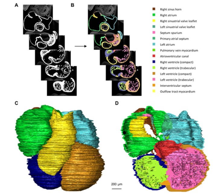
Quantified growth of the human embryonic heart
The size and growth patterns of the components of the human embryonic heart have remained largely undefined.
To provide these data, three-dimensional heart models were generated from immunohistochemically stained sections of ten human embryonic hearts ranging from Carnegie stage 10 to 23. Fifty-eight key structures were annotated and volumetrically assessed. Sizes of the septal foramina and atrioventricular canal opening were also measured. The heart grows exponentially throughout embr... Read more
Jaeike W. Faber, Jaco Hagoort, Antoon F. M. Moorman, Vincent M. Christoffels, Bjarke Jensen
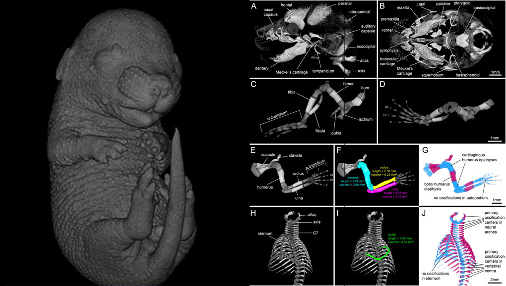
For decades, clearing and staining with Alcian Blue and Alizarin Red has been the gold standard to image vertebrate skeletal development. Here, we present an alternate approach to visualise bone and cartilage based on X-ray microCT imaging, which allows the collection of genuine 3D data of the entire developing skeleton at micron resolution.
Our novel protocol is based on ethanol fixation and staining with Ruthenium Red, and efficiently contrasts cartilage matrix, as demonstrated in wh... Read more
Simone Gabner, Peter Böck, Dieter Fink, Martin Glösmann, Stephan Handschuh
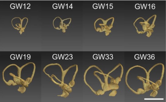
Progressive transformation of the otic placode into the functional inner ear during gestational development in humans leads to the acquisition of hearing perception via the cochlea and balance and spatial orientation via the vestibular organ.
Using a correlative approach involving micro-computerized tomography (micro-CT), transmission electron microscopy and histological techniques we were able to examine both the morphological and cellular changes associated with human inner ear devel... Read more
Lejo Johnson Chacko, David Wertjanz, Consolato Sergi, Jozsef Dudas, Natalie Fischer, Theresa Eberharter, Romed Hoermann, Rudolf Glueckert, Helga Fritsch, Helge Rask-Andersen, Anneliese Schrott-Fischer & Stephan Handschuh
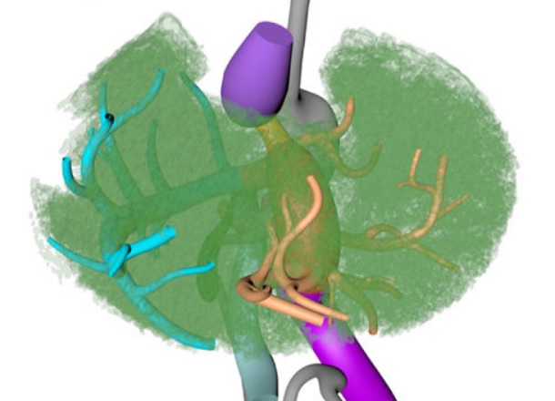
Couinaud based his well-known subdivision of the liver into (surgical) segments on the branching order of portal veins and the location of hepatic veins. However, both segment boundaries and number remain controversial due to an incomplete understanding of the role of liver lobes and vascular physiology on hepatic venous development. Human embryonic livers (5–10 weeks of development) were visualized with Amira 3D-reconstruction and Cinema 4D-remodeling software.
Read more
Jill P. J. M. Hikspoors, Mathijs M. J. P. Peeters, Nutmethee Kruepunga, Hayelom K. Mekonen, Greet M. C. Mommen, S. Eleonore Köhler & Wouter H. Lamers
The Academic Medical Center (AMC) uses Amira to build a 3D Atlas for Human Embryology
The 3D Atlas of Human Embryology project was funded by the Academic Medical Center (AMC) in Amsterdam, the Netherlands, in 2009. Since then, over 75 students, under the supervision of embryologists of the Department of Anatomy, Embryology & Physiology, have contributed to this labor-int... Read more
Academic Medical Center